Fresian horse
Male castrated
14 Years of age
Presented for acute severe lameness (graded 5/5) front right limb after training on the lunge.
Radiographs of the distal front right limb were taken.
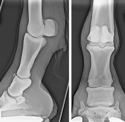
Lateromedial (on the left image) and dorsopalmar (on the right image) centred on the pastern region front right
Dorsomedial-palmarolateral (on the left image) and dorsolateral-palmaromedial (on the right image) oblique views centred on the pastern region front right.
Radiographic findings and diagnosis
- There is mild soft tissue swelling accentuated on the medial aspect of the limb, from the fetlock joint to the PIP joint.
- There are multiple, small, separated mineral opacities on the lateral aspect at the level and dorsal to the PIP joint. The biggest one (approximately 8mm in lenght, arrow) is lateropalmar and proximal to the PIP joint. Another one is dorsal at the level of the proximal P1. Multiple smaller fragments are also present dorsolateral to the joint.
- The medial border of the distal P1 is mildly irregular and of heterogenous opacity (arrowhead).
- The PIP joint is severely narrowed lateral (emtpy arrow).
- The radiographic diagnosis was: Fragment of dorsoproximal P1, suspicious of avulsion fragment of the lateral collateral ligaments of the PIP joint with joint instability and partial collapse. Severe desmitis or complete rupture of the collateral ligaments were suspected.
Radiographic examination
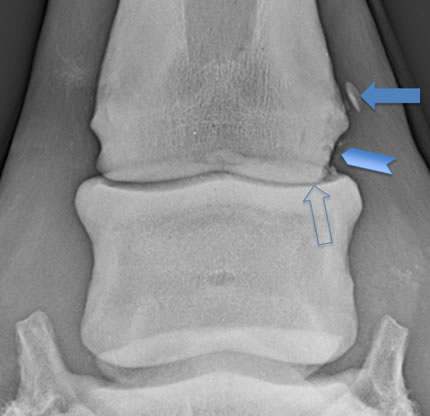
Close up of the DP view of the PIP joint front right
Comments
An additional stressed view was recommended.
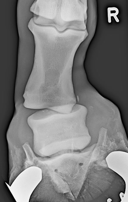
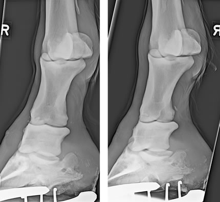
DP view of the distal front right limb stressed medially.
Comments
- The luxation of the PIP joint visible on the stressed view with severe widening medially and lateral displacement confirmed the joint instability.
- The horse was treated with an arthrodesis, the fragment dorsal to the proximal P1 was articular and was removed.
- Ruptured collateral ligaments may or may not be associated with avulsion fractures. If not, the radiographic diagnosis may depend on the stress radiography.

Lateromedial (on the left) and dorsopalmar (on the right) post OP view of the right front PIP joint.